News
Advanced tumour models reveal how glioblastoma weakens the immune response
LIH researchers use patient-derived 3D models to uncover how brain tumour cells disrupt T-cell activity, supporting the future development of more personalised immunotherapies
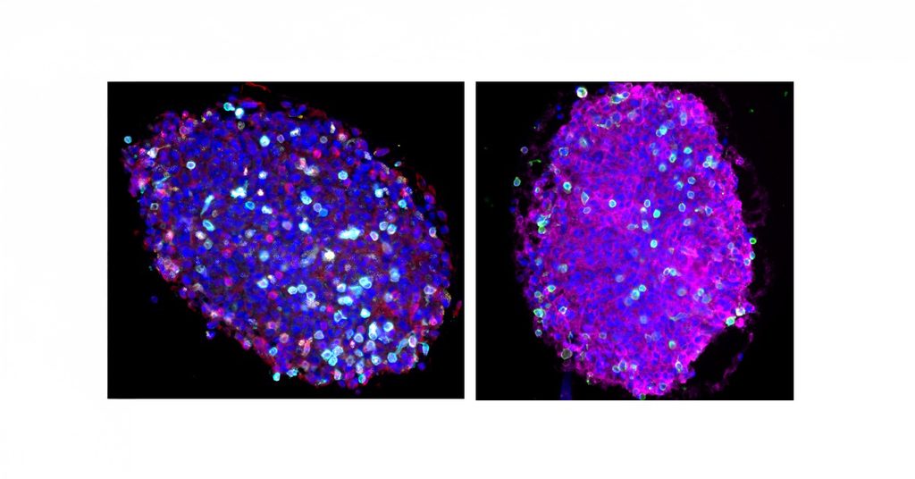
Researchers from the Luxembourg Institute of Health (LIH) have uncovered new insights into how glioblastoma cells suppress the immune response. Using advanced three-dimensional tumour models derived from patients, the study shows that direct contact with cancer cells can drive T cells towards dysfunctional and immunosuppressive states. Published in the Journal of Neuroinflammation, the findings also provide a patient-relevant platform for evaluating future immunotherapy strategies.
Glioblastoma is the most aggressive primary brain tumour in adults and remains extremely difficult to treat. Although immunotherapies have transformed care for several cancers, they have shown limited effectiveness against glioblastoma, partly because of the strongly immunosuppressive environment created within these tumours. Led by the NORLUX Neuro-Oncology Laboratory at the LIH Department of Cancer Research (DoCR), the team developed an advanced co-culture system combining patient-derived glioblastoma organoids with human T cells. These miniature 3D tumour models preserve important characteristics and cellular diversity of the original tumour. The models originated from the PRECISION-PDX collection, a Luxembourg resource developed from donated patient tumour tissue. By introducing T cells from patients with glioblastoma or healthy donors, the researchers could observe how immune cells entered the organoids and responded to tumour-derived signals.
Our organoid models allow us to examine the interactions between tumour and immune cells in a controlled but highly patient-relevant setting. They give us a more precise view of the local signals that weaken immune responses in glioblastoma and provide a valuable platform for investigating how these mechanisms might be overcome
explains Dr Anna Golebiewska, leader of the NORLUX Neuro-Oncology Laboratory and corresponding author of the study.
The study also addressed a debated question: whether glioblastoma cells escape immune surveillance by losing MHC class I molecules from their surface. These molecules help T cells identify abnormal or potentially dangerous cells. The researchers found that MHC-I remained detectable across patient tumours and diverse patient-derived models, including when tumour cells came into contact with T cells. This suggests that losing MHC-I is not the predominant way in which glioblastoma evades immune attack. Instead, direct contact with tumour cells appeared to alter T-cell behaviour. T cells that entered the organoids showed enhanced immunosuppressive traits, as well as diverse forms of dysfunction and exhaustion. By contrast, T cells exposed only to soluble substances released by the tumour were largely unaffected. The results therefore point to direct cell-to-cell interactions as an important driver of local immunosuppression.
The organoid platform provides a powerful system for studying how individual glioblastomas influence immune cells. In the future, it could help researchers evaluate potential immunotherapies, identify promising treatment combinations and explore why responses differ between patients.
The study brought together several LIH research groups and was conducted in collaboration with the Royal College of Surgeons in Ireland. The PRECISION-PDX collection was established with the participation of patients, the Neurosurgery Department of the Centre Hospitalier de Luxembourg, NORLUX and LIH’s Clinical and Epidemiological Investigation Center. The work received support from LIH, Télévie-FNRS, ERA-NET TRANSCAN-3, the Luxembourg National Research Fund (FNR), Fondation Cancer Luxembourg, Fondation du Pélican de Mie et Pierre Hippert-Faber under the Aegis of Fondation de Luxembourg and the European Union’s Horizon Europe programme.
The study was published in September 2026 in the Journal of Neuroinflammation with the full title “Advanced 3D patient-derived glioblastoma organoid-immune cell co-cultures reveal preserved MHC-I tumor cell surface expression and tumor cell-driven T-cell dysfunction”.
Scientific Contact
Related News